Total Pageviews

Showing posts with label Metabolic. Show all posts

Saturday, 9 June 2012

Refsum's disease

- autosomal recessive disorder
- caused by defective alpha oxidation of phytanic acid leading to its accumulation in tissues
- onset normally in late teens or 20s
- characterised by sensorimotor peripheral neuropathy, sensorineural deafness, anosmia, cerebellar ataxia, pes cavus, night blindness (retinitis pigmentosa), cardiac conduction abnormalities, epiphyseal dysplasia (shortening of 4th toe)
- elevated serum phytanic acid level
- treatment: dietary restriction of foods containing phytanic acid (chlorophyll)

Tuesday, 28 February 2012

Alkaptonuria

- rare (1:200,000) disorder of tyrosine metabolism
- autosomal recessive
- deficiency of homogentisic acid oxidase
- leads to excretion of large amount of homogentisic acid in urine and accumulation of oxidised homogentisic acid pigment in connective tissues (ochronosis)
- presents with dark urine, foci of gray-brown scleral pigment, darkening of concha, anthelic and helix of ear
- ochronotic arthritis is heralded by pain, stiffness and some limitation of motion of hips, knees and shoulders
- interverterbral disc calcification is characteristic of alkaptonuria and is seen on spinal X rays
- degenerative cardiovascular disease is increased in older patients
- treatment: Nitisinone


Sunday, 15 January 2012

Homocystinuria

- autosomal recessive
- reduced activity of cystathionine B-synthase
- accumulation of homocysteine and methionine interfere with collagen cross-linking
- similar to Marfan's syndrome, but with downward lens dislocation, venous and arterial thrombosis, developmental and mental retardation
- premature cardiovascular risk increased in heterozygote
- cyanide-nitroprusside test showed elevated urinary homocysteine
- management: methionine restriction, cysteine and pyridoxine supplements

Phenylketonuria

- autosomal recessive
- mutation of phenylalanine hydroxylase (chromosome 12)
- incidence 1:10,000
- inability to convert phenylalanine to tyrosine, results in hyperphenylalaninaemia and increased excretion of its metabolite (phenylketone) in urine
- presents with infantile spasm, developmental delay, musty smelling, pale skin, fair haired and blue eyed, eczema
- Guthrie screening test and measure level of phenylalanine by spectrofluorometric methods
- management: restriction of dietary phenylalanine with tyrosine supplement

Thursday, 12 January 2012

Mucopolysaccharidosis I and II (Hunter's and Hurler's)

- MPS are group of metabolic disorder caused by absence or malfunctioning of lysosomal enzymes needed to break down glycosaminoglycans (lysosomal storage disorder)

MPS type I (Hurler's syndrome)
- autosomal recessive
- cloudy cornea
- severe mental retardation
- gibbus
- coarse facies
- hepatosplenomegaly

MPS type II (Hunter's syndrome)
- X-linked
- clear cornea
- scoliosis, no gibbus
- less severe mental retardation

Sunday, 27 November 2011

Amyloidosis

- disorder of protein metabolism in which there is an extracellular deposition of pathological insoluble fibrillar proteins in organs and tissues
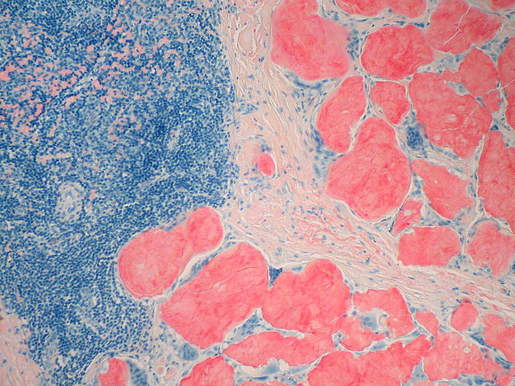
- diagnosis made by biopsy of affected tissue and positive Congo Red staining with red green birefringence under polarised light microscopy. Rectum or subcutaneous fat are relatively non-invasive site for biopsy

Primary amyloidosis (AL amyloidosis)
- in lymphoproliferative disorder (myeloma, MGUS, non-Hodgkin lymphoma), clonal plasma cells in bone marrow produce immunoglobulins that are amyloidogenic
- presents with nephrotic syndrome, cardiomyopathy (avoid digoxin), autonomic and sensory neuropathies, hepatomegaly, periorbital purpura and macroglossia
- echo : 'sparkling' appearance (restrictive cardiomyopathy)

Familial amyloidosis (transthyretin-associated)
- autosomal dominant
- renal disease less prevalent than AL amyloidosis and macroglossia does not occur

Reactive amyloidosis (AA amyloidosis)
- formed from serum amyloid A (SAA), which is an acute phase protein
- occurs in chronic inflammatory disorder
- presents with nephrotic syndrome, hepatomegaly and splenomegaly, cardiac involvement is much less common


Wednesday, 9 November 2011

Gaucher's disease

- deficiency in glucocerebrosidase
- results in accumulation of glucosylceramide in lysosomes of the reticuloendothelial system
- most common mutation is single base change causing substitution of arginine for serine
- characteristic pigmentation on exposed parts (forehand and hands), hepatosplenomegaly, pathological fracture
- diagnosis by finding a deficiency of lysosomal B glucocerebrosidase in leucocytes
- 3 types
  Type 1 (non-neuropathic) - commonest
  Type 2 (acute infantile neuropathic)
  Type 3 (chronic neuropathic)
- high ALP, ACE and Ig levels
- raised acid phosphatase
- definitive diagnosis made by genetic testing
- treatment: enzyme replacement treatment

Sunday, 6 November 2011

Familial hypercholesterolemia

- autosomal dominant (mutation on short arm of chromosome 19)
- heterozygote 1:500; homozygote 1:1 million
- mutations in LDL receptor or Apo B
- tendon xanthomata is hallmark of the disease
- elevated LDL, with normal Tg
- premature cardiovascular disease
- treatment: statins

File:Multiple hand xanthomas 18 yo case report.jpg

Wednesday, 12 October 2011

Hemochromatosis

- autosomal recessive
- commonest mutation is C282Y, in chromosome 6, affecting HFE gene
- most common single gene disorder in caucasians (carrier frequency 1 in 10)
- serum iron is elevated (>300mg/dL)
- serum transferrin saturation > 50% (most sensitive biochemical marker of iron overload)
- serum ferritin increased
- urinary iron excretion markedly increased (>2mg/24hour) by chelating drug desferioxamine
- diagnosis confirmed by DNA analysis
- presents with skin pigmentation (iron deposition stimulates increase melanin production), hepatic dysfunction, diabetes (bronze diabetes), hypogonadism, arthropathy (pseudogout), restrictive cardiomyopathy
- treatment: regular venesection (monitor by serum ferritin) - cardiomyopathy may improve with venesection

Tuesday, 11 October 2011

Wilson's disease

- toxic accumulation of copper in the liver and CNS (basal ganglia) due to failure of biliary copper excretion
- autosomal recessive
- defective gene ATP 7B (chromosome 13)
- prevalence 3:100,000
- normal copper absorption but defetive intrahepatic formation of caeruloplasmin
- features
   - onset in childhood/adolescence
   - hepatic dysfunction (cirrhosis)
   - CNS involvement (chorea, oromandibular dystonia, tremor, seizure, cerebellar signs, psychosis)
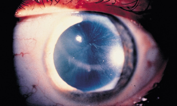
   - Kayser-Fleischer ring (copper deposition in Descement membrane)
   - hemolysis
   - Fanconi syndrome
   - low serum uric acid
- diagnosis
   - decrease in serum caeruloplasmin
   - increase in hepatic copper content
   - increase urinary excretion of copper (further increase following administration of penicillamine)
- treatment: copper chelators (penicillamine and trientine), zinc

Wilson disease

Wednesday, 7 September 2011

Porphyria

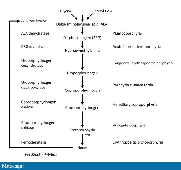
- Porphyrias are group of inherited disease resulting from deficiencies in haem biosynthetic pathway
- Acute porphyrias : acute intermittent porphyria, variegate porphyria, hereditary coproporphyria
- Non acute porphyria : cutaneous hepatic porphyria, congenital porphyria, erythropoeitic protoporphyria
- precipitants of acute attacks are: stress, infection, pregnancy, menstruation, starvation, drugs (sulphonamides, barbiturates, phenytoin)
- Haematin can help as it acts as negative feedback operator on the porphyrin synthetic pathway
- enzyme inducer exacerbate porphyria as they induce the pathway
- Ehrlich's test: add one volume of Ehrlich reagent to one volume of urine and urine turns red. Add two volumes of chloroform and red colour stays in upper layer
- Variegate porphyria is associated with raised urinary & fecal protoporphyrins
- Hereditary coproporphyria is associated with raised urinary & fecal coproporphyrins

Essential MRCP facts about porphyria
- All porphyrias have autosomal dominant inheritance except congenital porphyria
- Only acute porphyrias develop neurological consequence (ALA is neurotoxic)
- All porphyrias are photosensitive, except acute intermittent porphyria
- If the name sound's "inherited" eg.congenital porphyria or hereditary coproporphyria, then it is extremely rare.


Porphyria cutanea tarda
- most common subtype of porphyria
- deficient activity of enzyme uroporphyrinogen decarboxylase, causing accumulation of uroporphyrinogen III
- most common presenting sign is fragility of sunexposed skin after mechanical trauma, leading to erosion and bullae on dorsal hand, forearm and face
- diagnosis is by increased plasma and urinary porphyrins
- treatment: chloroquine, venesection

The hand of a patient with VP showing skin lesions

Sunday, 4 September 2011

Cystinuria

- autosomal recessive
- defective tubular reabsorption and jejunal absorption of cysteine and dibasic amino acids (COAL - cysteine, ornithine, arginine, lysine)
- commonest cause of renal stone in children where a metabolic cause is identified
- presents with chronic backache, recurrent urinary stones
- cysteine is highly insoluble at acid pH and form radio-opaque calculi
- urine FEME: pathognomonic hexagonal crystal

- management: large fluid intake, alkalinisation of urine and penicillamine / tiopronin /captopril

Sunday, 28 August 2011

Fabry's disease

- X-linked recessive lysosomal storage disorder
- deficiency of alpha-galactosidase A , causing accumulation of globotriaosylceramide
- myelin deposits in tubular epithelium and vascular endothelium resulting ischemic nephropathy
- presents with peripheral neuropathy (burning sensation of extremities evoked by vigorous exercise, hot weather or febrile illness) and cardiac conduction defect
- Absent of alpha-gal A in leukocytes confirm the diagnosis
- slit lamp examination revealed microscopic lipid deposits (corneal verticillata)
- skin lesion is known as angiokeratoma corporis diffusum (most prominent periumbilically)